Content Guides and Sample Questions
Last verified on April 25, 2025
On this page
PLEASE NOTE: List of Constants and Physical Values for Use on the Part 1 Physics Exam
The ABR provides candidates with a list of constants, physical values, and related information, which can be found on this page. While the list includes many constants and physical values, the ABR does not warrant the list as a compilation of all constants and physical values needed on the exams. Candidates should review the list carefully before their exams to familiarize themselves with the contents and list organization.
Content Guides
The content of all ABR exams is determined by a panel of experts who select the items based on a content guide that the ABR publishes. The content guides are assembled using guidance from medical physics organizations. The content guides are general documents, and individual exam items may not appear to be exactly congruent with the content listed in the guide. In addition, since there is only a limited number of items on any exam, selected items will only be a sample from the larger domain of the content guide. To understand how exam questions are written and learn more about different types of exam questions, please see the ABR Item Writers’ Guide. For a look at the extensive QA process that each question goes through, please see the Illustrated Life Cycle of an ABR Exam Item. For information about the remote exam delivery format and what to expect on exam day, please see the Medical Physics Remote Qualifying Part 1 Exam Guide.Part 1: General Content Guide
- Atomic / Nuclear Physics, Sources of Radiation, Interaction of Radiation with Matter
- Basic atomic and nuclear physics
- Radioactivity
- Sources of radioactive material
- Radioactive material uses and safety
- Radiation generating equipment: photons, electrons and heavy particles
- Interactions of photon and particle radiation with matter
- Dosimetry concepts and units
- Spatial distribution/transmission of radiation (photons, protons and electrons
- Radiation Instrumentation and Measurement
- Gas-filled detectors
- Scintillation detectors
- Solid state detectors
- Neutron detectors
- Emerging and miscellaneous detectors
- Measurement procedures
- Quality control and quality assurance
- Applications in imaging, nuclear medicine, therapy and safety
- Diagnostic Medical Physics
- Radiography, Fluoroscopy and Mammography
- Computed Tomography
- Ultrasound
- Magnetic Resonance
- Modality comparison, image features and artifacts
- Endogenous and exogenous contrast
- Modality facility considerations, safety
- Methods of quality control and quality assurance
- Nuclear Medical Physics
- Scintillation cameras
- Image acquisition and reconstruction
- Common radionuclides
- SNR, subject/image contrast
- Spatial resolution
- Mechanical aspects: accuracy, precision
- Single Photon Emission Computed Tomography (SPECT)
- Positron Emission Tomography (PET)
- Modality comparison, image features and artifacts
- Image processing and analysis
- Applications, dose, facilities and safety
- Hybrid imaging (SPECT/CT, PET/CT, PET/MR)
- Methods of quality control and quality assurance
- Counting principles
- Physical, biological, and effective half-life
- Therapeutic Medical Physics
- Clinical linear accelerator principles, collimation and mechanical aspects
- Clinical kV and MV photon beam characteristics
- Clinical megavoltage electron beam characteristics
- Clinical proton beam characteristics
- Comparison of clinical photon, electron and proton beams
- Dose functions: PDD, TAR, TPR, TMR, SMR
- Principles of radiation treatment planning
- Basic dose (monitor unit) calculation
- Brachytherapy
- Radiation safety and protection, patients and personnel
- Methods of quality control and quality assurance
- Radiation Protection, Safety, Professionalism and Ethics
- Principles of radiation safety
- Radiation risk and epidemiological data
- Radiation protection regulations: NRC and Agreement States
- Radiation areas
- Regulatory exposure limits
- Radiation protection program
- Radioactive source management and security
- Transportation of radioactive materials
- Shielding design for diagnostic, nuclear medicine and therapeutic installations
- Signage for diagnostic, nuclear medicine and therapeutic installations
- Nonionizing radiation safety
- Mechanical and electrical safety
- Principles of quality assurance and quality control
- Management of radiation accidents and large-scale radiological events
- Professionalism and ethics
- Informatics, Mathematics, Statistics, Image Processing & Analysis
- Mathematics relevant to medical physics
- Statistics and biostatistics
- Medical image analysis and processing
- Observer performance and ROC analysis
- Informatics
Part 1: Clinical Content Guide
- Anatomy
- Breast
- Cardiovascular
- Digestive System
- Musculoskeletal
- Neurological System
- Reproductive/Endocrine
- Thoracic Cavity
- Urinary System
- Lymphatic System
- Radiation Biology
- Physics and chemistry of radiation interactions with matter
- Molecular and cellular radiobiology
- Tumor radiotherapy
- Normal tissue response to radiotherapy
- Time dose fractionation
- Radiobiological basis of radiation protection
- Radiation accidents and environmental radiation exposure
- Diagnosis and medical management of radiation syndromes
- Deterministic effects
- Stochastic Effects
- Radiation carcinogenesis
- Heritable radiation effects
- Effects on the developing embryo
- Human Physiology
- Nervous system
- Musculoskeletal system consists of the
- Cardiovascular system
- Respiratory system
- Digestive system
- Integumentary system
- Urinary system
- Reproductive system
- Immune system
- Endocrine system
- General Medical / Radiology / Radiation Therapy Terminology
- Medical Root Words
- Diagnostic Radiology terminology
- Radiation Therapy terminology
- Clinical Procedure Applications
- Diagnostic Radiology
- Radiation Therapy
- Pathology
- Neoplastic Diseases
- Benign Disease
- Infectious Diseases
- Congenital and hereditary diseases
- Inflammatory
- Trauma
- Cardiovascular Diseases
- Neurological
Sample Questions
Part 1: General Sample Questions
(includes new item types)- Beyond the depth of maximum dose, what is the relative behavior of dose and kerma?
- Dose and kerma fall off equally.
- Kerma falls off faster than dose.
- Dose falls off faster than kerma.
- Dose falls while kerma rises.
- Dose rises while kerma falls.
- At 75oF and 1 atm, a vented ionization chamber should have its reading “temperature corrected” by multiplying the reading by which factor? (Assume the ionization chamber has a calibration factor from an AAPM-accredited U.S. calibration laboratory.)
- 0.994
- 1.003
- 1.006
- 1.021
- The mass attenuation coefficient of bone (density of 1.8 g/cm3) is 0.2 cm2/g for an 80-KeV gamma ray. What percentage of 80-KeV photons is attenuated by a slab of bone 4 cm thick under conditions of narrow beam geometry?
- 36%
- 45%
- 55%
- 64%
- 76%
- What is the fraction of the initial number of radioactive atoms remaining after a period of time t?
- λ
- λt
- -λt
- e-λt
- 1-e-λt
- Reduced contrast in a therapy verification image compared with a simulator radiographic image is primarily a result of which process?
- Increased number of pair productions
- Increased number of Compton interactions
- Increased number of photoelectric interactions
- Decreased number of photoelectric interactions
- Decreased number of Compton interactions
- A 2 cm3 ionization chamber is placed in a radiation field of 100 R/s. The current generated is how many amperes?
- 5.1 x 10⁻¹¹ amperes
- 6.3 x 10⁻¹⁰ amperes
- 5.1 x 10⁻⁹ amperes
- 6.3 x 10⁻⁸ amperes
- 5.1 x 10⁻⁷ amperes
- At a radiologist’s workstation, the instantaneous exposure rate through the wall from a chest image receptor is 500 mR/h during exposures of 10 mAs at 100 kV and 100 mA. For 200 exposures per week, what is the approximate maximum exposure at the workstation?
- 0.28 mR
- 2.8 mR
- 28 mR
- 280 mR
- 2800 mR
- When the energy of incident x-ray photons just exceeds the binding energy of inner shell electron orbital, what is the abrupt increase in the attenuation coefficient called?
- Compton edge
- Absorption edge
- Photopeak
- Characteristic transmission
- Characteristic disintegration
- Ten 1024 x 1024 digital images with 64 shades of gray are to be transmitted at the rate of 1.049 x 105 bits/s. Approximately how long will it take to complete the transmission?
- 10 seconds
- 30 seconds
- 1 minute
- 10 minutes
- 1 hour
- A group of patients exhibits lifetimes that are normally distributed with a mean of 36 months and a standard deviation of 3.0 months. What percentage of these patients will survive at least 42 months?
- 2.5%
- 5.0%
- 33.0%
- 67.0%
- 95.0%
Answers for this section:
1. A
2. C
3. E
4. D
5. D
6. D
7. B
8. B
9. D
10. A
New Item Types
Fill-in-the-blank
The candidate must type in the correct response:
- For a pressure of 756 mm Hg and a temperature of 21°C, the temperature-pressure correction factor for an unsealed ion chamber is ____________. (Round to the third decimal place.)
Answer: 1.002
How to get to the answer: ((273.15 + 21)/295.15) x 760/756 = 1.0019. Round to third decimal = 1.002
Answer: B and C
Case-based Items
There will be a block between the questions that permits the candidate to review the previous question, but not to change the answer.
Case Number 1
- For a 2D ultrasound image that consists of 100 A-mode lines resolving an object at a depth of 15 cm and an acquisition time per scan line of 13 µs/cm, what is the total time per frame?
- 5.0 ms
- 11.2 ms
- 19.5 ms
- 27.3 ms
BLOCK
- If the time per frame is 19.5 ms, what is the frame rate?
- 26 Hz
- 51 Hz
- 100 Hz
- 769 Hz
- 5000 Hz
Answer: 1 = C; Answer 2 = B
Case Number 2
- Eighty-five percent of an injection of 99mTc sulfur colloid is cleared from the blood by the liver. What is the time-integrated activity coefficient in the liver? (Assume a biological half-life of 3 hours.)
- 1.4 hours
- 2.5 hours
- 3.1 hours
- 4.3 hours
Comments: Lamda_eff = 0.693/3 h + 0.693/6 h = 0.3465/h (2 h)
0.85*2 h/0.693=2.5 h
BLOCK
- Given an administered activity of 2.8 mCi, a time-integrated activity coefficient of 2.5 hours, and an S factor [liver→kidney] of 3.9 × 10⁻⁶ rad/μCi-h, what is the absorbed dose to the kidney from the liver?
- 18 mrad
- 21 mrad
- 27 mrad
- 34 mrad
Comments: 2800 uCi*2.5 h*3.9 x 10^-6 rad/uCi-h = 27 mrad
Answer: 1 = B; Answer 2 = C
Part 1: Clinical Sample Questions
- The human typically has how many cervical vertebrae?
- 12
- 10
- 7
- 5
- 4
- A fetus receives a dose of 2 Gy during weeks 20 to 39 of pregnancy. After birth, the child has an increased risk for what condition?
- Trisomy 21
- Leukemia
- Microcephaly
- Neonatal death
- A patient is suspected of having kidney stones. What is the most appropriate exam?
- CT
- MRI
- Abdominal radiography
- Endoscopic retrograde cholangiopancreatography
- Hysterosalpingogram
- What is the most common location for cancer in the breast?
- Lower inner quadrant
- Lower outer quadrant
- Upper inner quadrant
- Upper outer quadrant
- For ionizing radiation, how does the OER vary as LET increases from 1 to 100 KeV/μm?
- Increases
- Decreases
- Remains the same
- Increases then decreases
- Decreases then increases
Answers for this section:
1. C
2. B
3. A
4. D
5. B
New Item Types
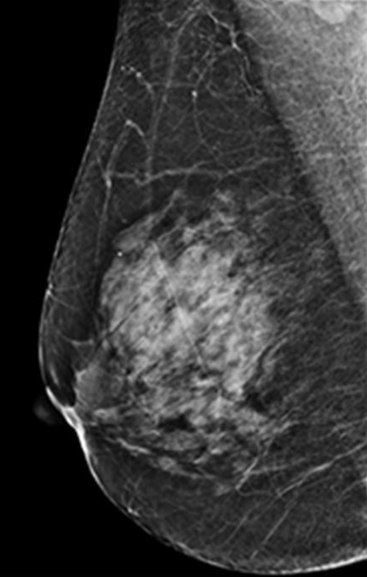
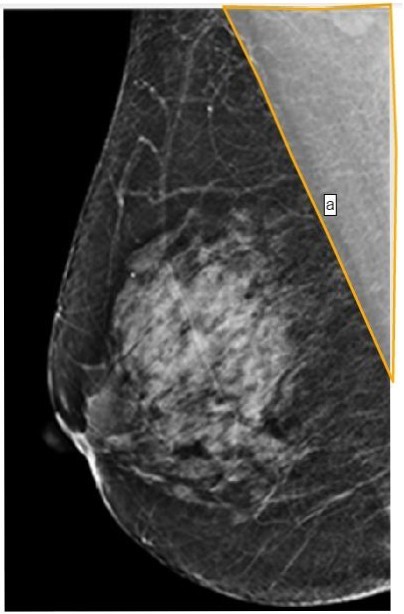
Point-and-Click
The candidate must identify a region on an image.
-
- Point and click on the pectoralis muscle.